Special Senses

Unit 13 Synopsis
In this unit, we will examine the various special senses including gustation, audition, equilibrium, vision, and somatosensation.
What are the special senses?
Gustation
Sense of taste
Vision
Sense of sight
Audition
Sense of hearing
Blephar-
Eyelid
Orbit-
Eye socket
AFFIXES
Tympan-
drum
Macula-
Spot
Ocul-
Eye
Ophthalm-
Eye
Olfact-
Smell
-lith
Stone
Aqua-
Water
rhino-
nose
Med-
Middle
Ot(o)-
ear
Gust-
Taste
Ossi-
Bone
Gustation - Tastes
Sweet: perception of monosaccharides
Salty: perception of sodium cations [Na+]
Sour: perception of hydrogen cations [H+], which determine pH
Bitter: wide variety of receptors, useful for protection. Stimulates gag reflex to avoid poison
Umami (savory): receptor for L-glutamate (amino acid); commonly received in protein rich food
Possible sixth taste sense specific to lipids

Gustation Anatomy
Primary structure is the tongue
Surface is lined with stratified squamous epithelium
Papillae: bumps on the tongue
Taste Buds: sensory organ of the tongue; found within the papillae
Gustatory epithelial cells: taste receptor cells
Basal epithelial cells: stem cells that make new gustatory epithelial cells

There are four types of papilla:
Fungiform
Filiform
Foliate
Circumvallate
Audition
Sense of hearing.
Predominant sensory organ is the ear made of 3 parts
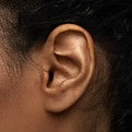
The External Ear
Pinna (auricle): the cartilage area that "catches" the sound waves to bring it into the ear
External auditory meatus: the auditory canal
Tympanic Membrane: eardrum that is the connective tissue boundary between external and middle ear

The Middle Ear
Tympanic membrane: relay center between the outer and inner ear
Auditory ossicles: malleus, incus, stapes that function to amplify the sound waves
Oval Window: the auditory ossicles conduct vibrations to the oval window to get the inner ear fluid moving
Tiny synovial joints and ligaments within the ossicles reflexively contract to minimize damage from loud sounds
Eustachian (yüˈstāshən) tube: passage from the middle ear to the pharynx that aids in equalizing pressure around the eardrum

The Inner Ear
The bony labyrinth
Functions to turn the physical vibrations into action potentials that the brain can interpret.
Vestibule: structure that helps maintain balance
Semicircular canal: aid in maintaining balance when the head rotates
Cochlea: contains hair cells that vibrate at different frequencies that will stimulate the organ of Corti
An action potential is then sent through the Cochlear nerve to the auditory cortex of the brain
Key Terms to Know

Receptors
Exteroceptor
sensory receptor positioned to interpret stimuli from the external environment, such as photoreceptors
Mechanoreceptor
receptor to physical distortion
Chemoreceptor
receptor detects chemical, such as in taste and smell
Interoceptor
receptor that interprets stimuli from internal organs
Nociceptor
receptor of pain stimuli
Proprioceptor
receptor detects physical location